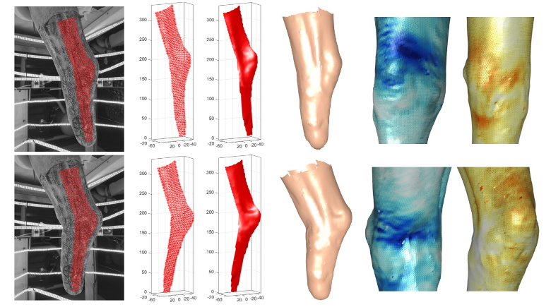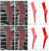
Effective prosthetic socket design following lower-limb amputation depends upon the accurate characterization of the shape of the residual limb as well as its volume and shape fluctuations. This study proposes a novel framework for the measurement and analysis of residual limb shape and deformation, using a high-resolution and low-cost imaging system. A multi-camera stereo system, featuring 21 Raspberry Pi camera modules, was designed and built to capture sets of simultaneous images of the entire residuum surface. Additionally, a custom three-dimensional calibration object was designed and 3D printed, which allowed for the instantaneous calibration of the system. The images were then analyzed using MultiDIC: a specially developed open source three-dimensional digital image correlation (3D-DIC) MATLAB toolbox, to obtain the accurate time-varying shapes as well as the full-field deformation and strain maps on the residuum skin surface. In-vivo measurements on one transtibial amputee residuum were obtained during sets of knee flexions, muscle contractions, and swelling upon socket removal. It was demonstrated that the new framework can be employed to quantify with high resolution the time-varying residuum shapes, deformations, and strains. In addition, the enclosed volumes and cross-sectional areas were computed and analyzed. This novel low-cost framework provides a promising solution for the in-vivo quantification of residuum shapes and strains, as well as the potential for characterizing the mechanical properties of the underlying soft tissues. These data may be used to inform data-driven computational algorithms for the design of prosthetic sockets, as well as of other wearable technologies which mechanically interface with the skin.

