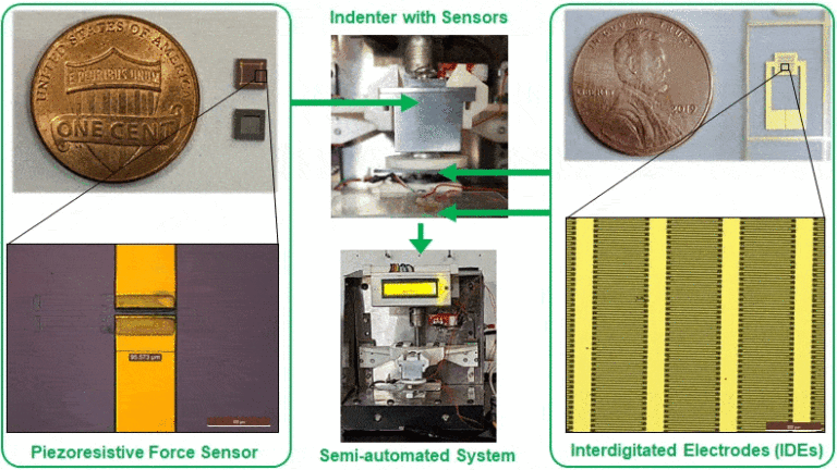
Accurate identification of surgical margins in brain tumors is of significant prognostic importance. Despite the availability of methods such as 5-ALA and image guidance, recognizing tumor boundaries is highly subjective and dependent on recognizing subtle changes in tissue characteristics, including texture and color to aid distinction. The study aims to design and develop a semi-automated system integrated with MEMS-based electromechanical sensors to arrive at an objective and reliable method of distinguishing tumors from normal brain tissue.
Microforce sensors and microchips with interdigitated electrodes (IDEs) were fabricated to characterize human brain tissues and tumors. These devices were packaged and integrated into a semi-automated system with a tissue indentation stage, data acquisition and processing unit, and an electrical impedance measurement unit. Simultaneous electrical impedance and viscoelastic characterization of three types of freshly excised gliomas (glioblastoma (GBM), astrocytoma (AST), and oligodendroglioma (OLI)) (N=8 each) and seventeen different normal brain regions (N=6 each) obtained postmortem was performed. The electrical impedance of gliomas was found to be significantly lower than corresponding normal regions. The difference in the impedance between individual tumor types and corresponding normal regions was also statistically significant, suggesting clear tumor delineation. There were distinct differences in the viscoelastic relaxation responses of high-grade and low-grade gliomas. White matter regions demonstrated higher impedance and faster stress relaxation compared to grey matter regions as a characteristic of their structural composition.
It is demonstrated that simultaneous electromechanical characterization of brain tumors and normal brain tissues can be an effective biomarker for tumor delineation, grading, and studying heterogeneity between the brain regions. The observations suggest the potential use of the technology in a clinical setting to achieve gross total resection and improve treatment outcomes by helping surgeons perform real-time risk evaluation during surgery.

