As the third main cause of disability worldwide, hearing impairment has been elevated to a major public health issue. Consequently, the World Health Organization promotes the development of
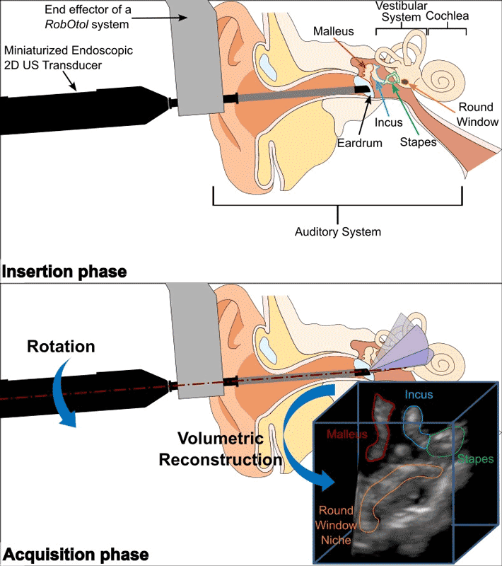
low-cost technologies for facilitating diagnosis. The aim of this study is two-fold: to demonstrate the feasibility of minimally invasive ultrasound (US) imaging of the auditory system using a miniaturized endoscopic 2D US transducer and to validate its clinical applicability to enable diagnosis and surgical navigation for otology in a safe way.
To this end, we introduced a novel probe that consists of a 18 MHz 24 elements curved array transducer with a distal diameter of 4 mm, which fits the external auditory canal (EAC). To obtain a 3D volume from acquired B-scans, rotation around the axis of the transducer was performed using a RobOtol system, a robotic platform dedicated for middle ear surgery, followed by a volumetric reconstruction procedure based on a scan-conversion algorithm. Since this imaging platform is meant for medical purposes, we focused on comparing the correctness of reconstructed volume’s geometry to the reality of acquisition. In the absence of any standard methodology, we proposed a new evaluation protocol that relies on the automatic registration and comparison of US volumes to a ground truth micro-CT acquisition of a set of wires as reference geometry. Additionally, we performed four volumetric acquisitions on a cadaveric head to explore the clinical applicability in a representative anatomical context.
Twelve acquisitions with different probe poses allowed to estimate a maximum error of reconstruction of 0.20 mm. Regarding the ex vivo acquisitions, volumetric reconstruction enabled the identification of critical structures of the ear such as the ossicles and the round window. This is the first time that volumetric US imaging of the human auditory system is performed without prior deterioration of the EAC.

