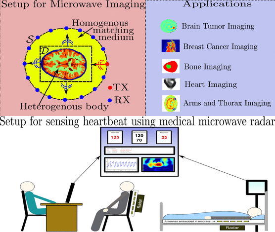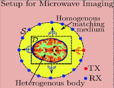
Widely used medical imaging systems in clinics currently rely on X-rays, magnetic resonance imaging, ultrasound, computed tomography, and positron emission tomography. The aforementioned technologies provide clinical data with a variety of resolution, implementation cost, and use complexity, where some of them rely on ionizing radiation. Microwave sensing and imaging (MSI) is an alternative method based on nonionizing electromagnetic (EM) signals operating over the frequency range covering hundreds of megahertz to tens of gigahertz. The advantages of using EM signals are low health risk, low cost implementation, low operational cost, ease of use, and user friendliness. Advancements made in microelectronics, material science, and embedded systems make it possible for miniaturization and integration into portable, handheld, mobile devices with networking capability. The fundamental notion of MSI is that it exploits the tissue-dependent dielectric contrast to reconstruct signals and images using radar-based or tomographic imaging techniques that can be quantitative or qualitative. Quantitative methods solve the ill-posed inverse EM problem through different iterative schemes to minimize the error between the measured and the modeled scattered field to obtain the electrical properties. Qualitative methods avoid solving ill-posed inverse problem by using radar-based techniques, and can be used in cases where the objective is the detection of the strong scatterer like the tumor. MSI has been studied for tumor detection, blood clot/stroke detection, heart imaging, bone imaging, cancer detection, and localization of in-body RF sources. The feasibility studies of using MSI in various applications show the potential of MSI as low health risk and low cost alternative to the currently used imaging modalities like MRI and CT-scan. However, MSI is still far from maturity as many open challenges remain that have to be addressed before MSI can be used in a clinical setting.

