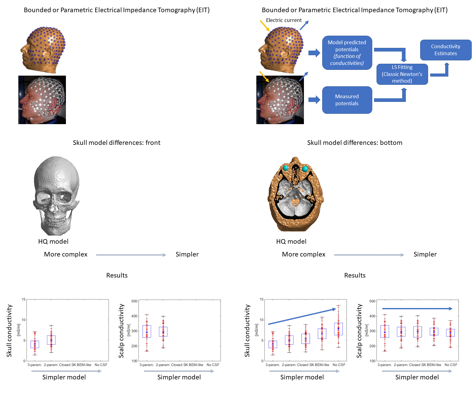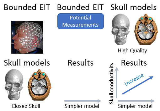
We estimate scalp, skull, compact bone and marrow bone electrical conductivity values using Electrical Impedance Tomography (EIT) measurements and assess the influence of skull modeling details on the estimates. We collected high density EIT data with 128- and 256-channel EEG electrode using 62 and 128 distinct current injection pairs, respectively, and built five 6-8 million finite element (FE) head models with different grades of skull simplifications for four subjects, including three whose head models serve as Atlases in the scientific literature (Colin27) or in commercial equipment (Philips Neuro/EGI’s GeoSource atlases). We estimated electrical conductivity of the scalp, skull, marrow bone and compact bone tissues for each current injection pair, each model, and each subject. We found that simplifications of the FE models, such as closing anatomical skull openings and omitting the CSF layer, produce an overestimation of the skull conductivity by 50-70%, as compared to the reference high-quality model. The average extracted tissue conductivities were found to be: 288±53 (the scalp), 4.3±0.08 (the compact bone), and 5.5±1.25 (the whole skull) mS/m. The marrow bone estimates showed large dispersion. Present EIT estimates for skull conductivity are lower than typical literature reference values, but the previous in-vivo EIT results are likely overestimated due to the use of simpler models like nested layer boundary element models. Typical literature values of 7-10 mS/m for skull conductivity should be adjusted to the present estimated values when using detailed skull head models. We also provide subject specific conductivity estimates for widely used Atlas head models. Incorporating the findings from EIT provides more accurate conductivity values as inputs for detailed head models and the use of EIT on individual subjects allows for individualized conductivity measurements, which should improve precision in applications such as electrical source localization and transcranial electrical stimulation.

