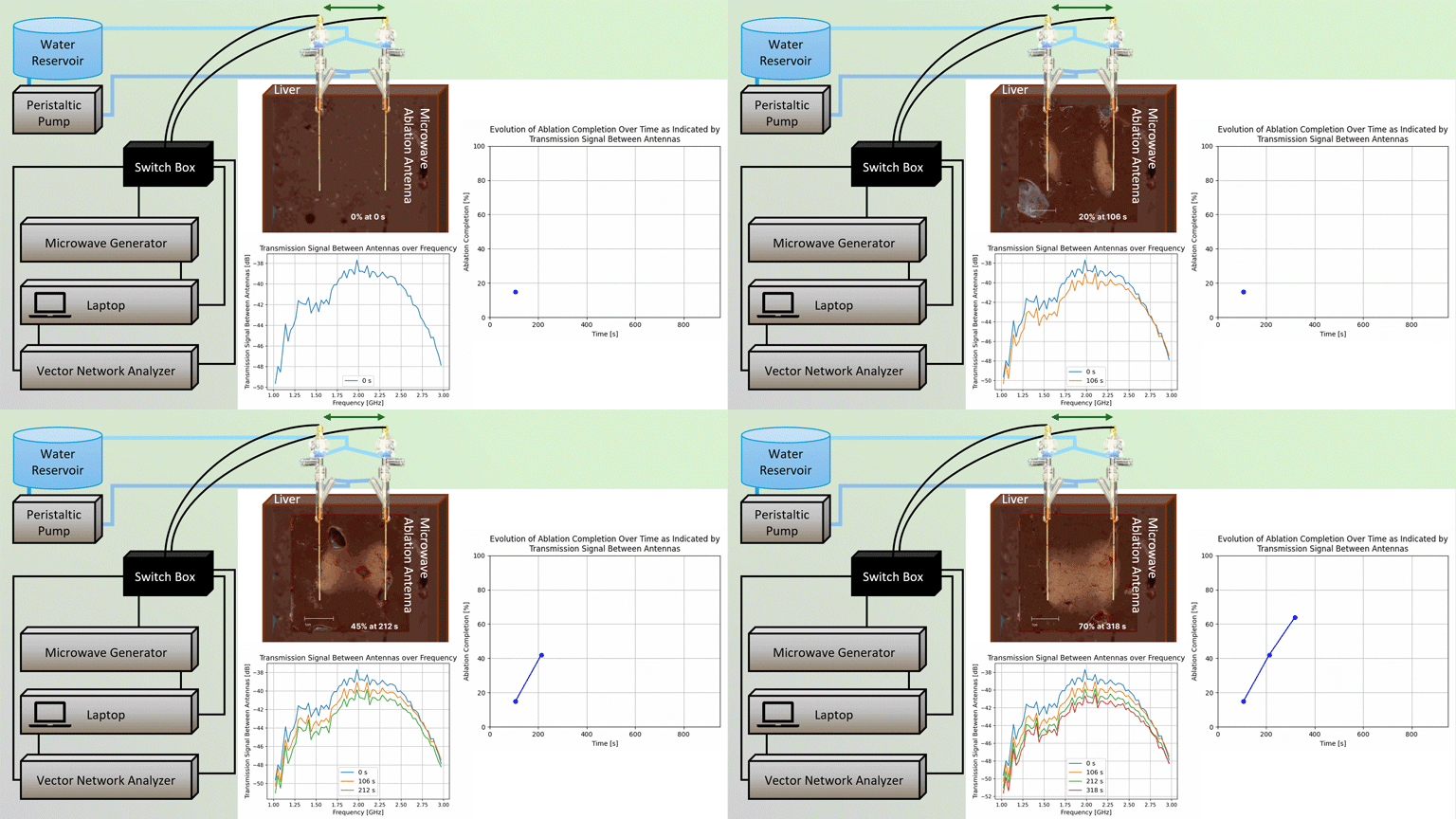
Microwave ablation (MWA) offers a minimally-invasive approach for image-guided treatment of localized tumors in the liver and other organs. During an ablation procedure, clinicians aim to balance the dual goals of achieving adequate treatment margins, while limiting damage to non-targeted tissue. A challenge is the limited intra-procedural feedback for monitoring transient evolution of the ablation zone and thus determining treatment endpoint. This study introduces a method for real-time monitoring of MWA, utilizing transient changes in broadband transmission signal spectra between two ablation antennas, to assess ablation zone progression. The presented method is experimentally evaluated in ex vivo liver tissue.
We designed and implemented a custom instrumentation system allowing for intermittent switching between ablation mode and assessment mode and tested the proposed system with a pair of directional MWA antennas in ex vivo liver tissue. Transmission signals were measured every minute for applied power levels of 30 – 45 W/antenna for 53 – 1219 s. At the conclusion of the experiment, an image of the cross-sectional ablation zone was captured. We (a) processed the cross-sectional images to calculate a ground truth of ablation completion, based on the ratio of ablated pixels between the two antennas and the anticipated extent of complete ablation, and (b) processed the broadband transmission signals to estimate ablation completion. We assessed the accuracy of predicting ablation zone completeness from the transmission signal data as compared to ground truth images.
The ablation zone prediction from the transmission signal method was within 5% of that observed on ground truth images. Ongoing studies are targeted towards evaluating the presented method’s performance in an in vivo large animal model. If successfully developed and translated, the presented system offers a means for leveraging ablation antennas for also monitoring progress of ablation procedures and determining treatment endpoint.

